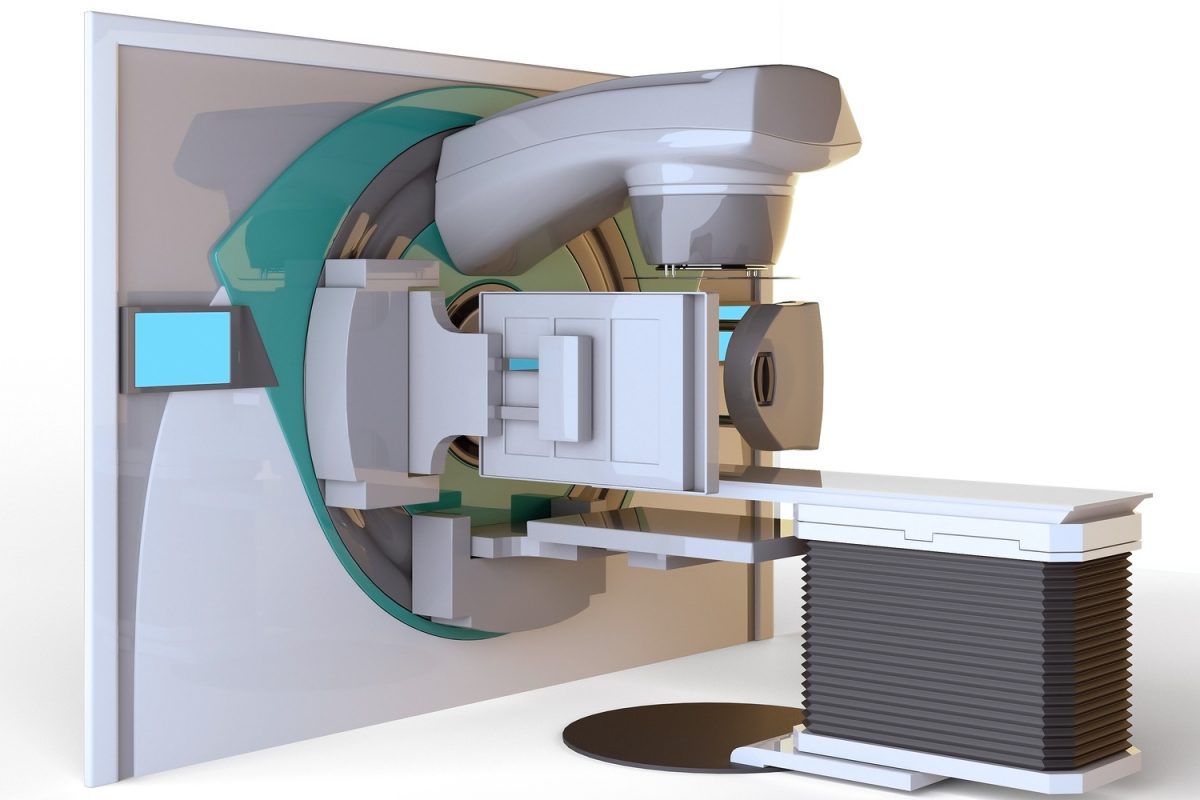
Magnetic Resonance Imaging (MRI) is an exciting tool to promote breast health and fight breast cancer. MRI combines the highest quality pictures of breast tissue with the highest cost of any breast imaging modality. The best for the most. Given the wide but somewhat confusing use of breast MRI, it is worth a moment to review this technology.
MRI uses magnetism to create images of tissue. There is no X-ray exposure. Ions in water and fat change in a high-powered magnetic field. Differences in tissue ions are detected by applying magnetic pulses. The pictures are in 3-D and show details of muscle, fat, supportive fibers, blood vessels and glands. MRI detects breast cancer by highlighting tiny blood vessels that grow in cancer.
The scan is taken in the prone (face down) position over a specialized MRI coil and takes about 45-60 minutes. Taking a mild sedative (like Xanax) before the test can help the claustrophobia experienced by many patients. Ideally, images should be taken between the 7th and 14th days of the menstrual cycle.
The test requires IV injection of a contrast agent called gadolinium (“gad”). This is not iodine based and allergies are uncommon. MRI contrast is not needed if the goal of the scan is to check a silicone breast prosthesis. However, allergy to gad and significant kidney disease are contraindications. Patients with pacemakers, pieces of metal or recent surgical clips can not have MRIs.
MRI is the most sensitive test for detection of breast cancer. However, there is no evidence that its greater ability to detect small cancers (more then a screening mammogram or ultrasound) results in increased cure rates. Therefore, as it is 10 times more expensive then mammography ($102- $212 vs. $2,000 – $6,200) and has an increased rate of false positives (sees things which need biopsy but do not turn out to be cancer) it is indicated only in select situations.
Breast MRI is recommended in addition to mammogram as a yearly or semi-yearly screening test in women that have a lifetime risk of breast cancer of 20 percent or more. This generally means women who carry the breast gene (BRCA 1/2) or have a strong family history of breast or ovarian cancer. Women who have a history of therapeutic chest radiation (such as in treatment of Hodgkin’s Disease) should start getting annual MRIs after age 25. In addition some experts advise breast MRI when mammograms are difficult to read (i.e. are “dense”).
MRI’s are commonly used to follow up inconclusive changes found on routine screening mammograms. The MRI can help decide if a biopsy is required or if the abnormality seen on the mammogram is likely to be benign. When needed, MRI can be used to guide a biopsy.
75 percent of women with a new diagnosis of breast cancer have an MRI of both breasts before surgery. The test can detect other breast cancers in the same breast, not felt by exam or not seen on mammogram. In addition, when a new breast cancer is found, there is an increased chance of breast cancer in the other breast. The use of MRI in this way is controversial, as there is a significant increase in unnecessary biopsies and mastectomies. MRI used in this way has not yet been proven to save lives.
Silicone implants can rupture and leak into the breast requiring removal and replacement of the implant. Breast MRI is quite effective for detecting rupture. MRI can also be used to differentiate false tumors caused by direct silicon breast injections (not done in many years) verses cancer. MRI cannot evaluate saline implants.
In conclusion, the use of breast MRI should be confined to high-risk situations where clear differences in treatment will be made based on the findings of the scan. MRI supplements the use of mammograms in breast cancer diagnosis and screening. This is not a “routine” test, but a special tool to evaluate particular problems.


6 Comments