Cancer 101: PET Scans

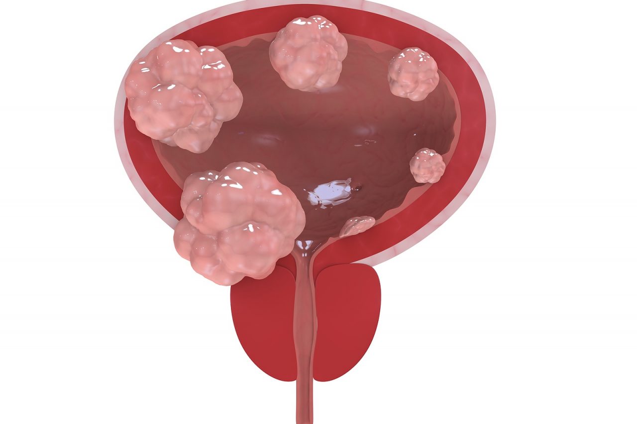
Let us play doctor.   Believe for a moment that you are sitting in your office advising a patient.   On the patient’s chest X-ray is a half-inch spot.  The patient is frightened and you are concerned. How you will determine if the spot is cancer or might it be a scar from an old infection?

Do you take the patient to surgery and remove the lesion?   Surgery requires anesthesia and causes pain.  An operation carries the risk of bleeding and infection.  Insert a needle?  That is hard to do in the middle of the lung (“Please hold your breath for 5 minutes.”).  This carries the risk of pneumothorax and bleeding.  Often needle biopsies miss.  Wait a couple months?  If it is cancer, it might grow.  What is the simplest, safest answer?  Order a PET Scan.

Positron Emission Tomography (PET Scan) cannot tell you the shape or the exact size of the spot.   It is not very good at determining location.  However, PET Scan can tell whether a lesion is growing.  PET is a test for tissue activity.  A scar is not very active and so the PET Scan does not see it.  PET does not detect most infections.  A cancer is rapidly expanding tissue and is “hot” on a PET Scan.

Cancers grow as dividing tissue require nutrients.  Cancers require sugar.  In order to perform PET Scans a particular sugar is manufactured. This sugar is radioactive.  Fluorine-18 flurodeoxyglucose, known at FDG, is the radioactive tracer used in PET Scans.  The patient receives this as an injection, getting a small radiation exposure, less than most CT Scans.   Cancer cells take up this FDG sugar and it is trapped inside. The PET Scanning machine then measures the radiation signal.

The more radiation the cancer cell takes up, the “hotter” it is on the scan.  A lesion that is hot may be cancer.  By matching the PET Scan to other tests (such as a CT Scan as in a combined PET-CT) it is possible to tell where a tumor is located, what it is touching and by how hot it is, how likely it is to be cancer.

PET Scans have several different uses.  First, to determine if a lesion might be cancer.  Like the patient above, a PET might avoid further testing of a benign lesion.

Second, PET Scans can detect the spread of cancer.  It is critical at the start of the cancer process to accurately “stage the patient.”  By knowing whether a cancer may have metastasized, the oncologist can design the proper treatment.

During therapy, PET Scans can determine if the treatment is killing the cancer.  For example with lymphomas, many experts believe that if a PET Scan is not normal after two treatments, then continuing that treatment will fail.  This allows an early change in therapy.

Finally, PET Scans are used to monitor patients who are in remission, to be certain that cancer has not returned.  For this reason in some cancers, PET Scans are repeated 6–12 months after finishing treatment.

Like all tests, PET Scans have their limits.  First, PET does not work for all cancers.  In general, PET Scans are most useful for rapidly growing cancers such as Hodgkin’s disease, aggressive lymphomas and lung cancer.  It may also be helpful in breast cancer, gynecologic cancers and some sarcomas.  It is of mixed benefit in colon cancer,  kidney and pancreatic cancer.  PET is useless in leukemia, which do not form solid masses.

PET Scans are sensitive, able to detect cancers that are more than one centimeter in size.  However, the scans are not very specific.  This means that it frequently will detect things that are not cancer.  A recent infection or injury may show up on PET.  This means that when a PET Scan does not show uptake (meaning it is not hot) there is unlikely to be cancer.  However, when a PET Scan detects uptake of the FDG tracer, it means that it might be cancer, but is not definite.

Like most new technologies, PET Scans are very expensive, often costing thousands of dollars.  Because of this financial burden, insurance companies put significant limits on their use. The doctor needs to prove there is clear research data supporting the use of a PET Scan and explain how the result will change therapy.

Should we all get an annual PET Scan?  Absolutely not.  Forgetting the cost and the unneeded radiation exposure, the reality is that more people would be hurt then helped.  PET Scans would find  “hot spots” in many people that are not cancer.  These people would have unnecessary tests and biopsies, with attendant complications.  The average person would be frightened by all these false positives and hurt by complex follow-up.

Researchers are studying how to improve PET Scans. Which cancers and when?  How to combine PET with other tests.  The use of tracers other than FDG. How to use PET Scans to better design and target therapy. These studies will transform and perfect PET.

PET Scans are a marvelous tool.  Carefully applied and interpreted, PET can answer hard questions in a relatively simple way.  Speaking as a doc who spent many years staring at spots on x-rays and trying to magically divine their intent, it almost feels like an unfair advantage.  However, this is one case where I will be happy to use the test to cheat.

10 Comments

  • sherri
    Please translate the following: There is no FDG avid enlarged lumph nodes in the neck. There are several subcentimeter bilateral level II nodes and a right-sided level III node with maximum SUV of 3.5, nonspecific. There is a mildly prominent FDG uptake in/around bilateral posterolateral nasopharyngeal wall in/adjacent to the area of the fossa rosenmuller with maximum suv of 6.5 on the right and 4.9 on the left. This is of uncertain etiology, although it could represent physiologic longus coli muscle uptake or reactive adenoid uptake or focal inflamation.
    • James Salwitz, MD
      All scans and tests need to be viewed in the context of the patient and the questions being asked. It is very hard to "translate" a complex image such as a PET without knowing the medical history and without old scans if they were done. Having said this limitation, this scan does not show clear evidence of any medical problem. jus
      • sherri
        I'm sorry I wasn't more thorough. I had a CT scan of the lungs and there were some small nodules in the right lobe, in 1/2015. Repeated in 1/2016 and there was a change in one of two small nodules in right lobe. It had grown. Repeated CT scan 6/26/2016. There was a new nodule in the left lower lobe, so referred to Pulmonary dr. He ordered a PET scan. He is comfortable with rechecking the lungs in 3 months, but has referred me to an ENT. My initial question are the findings of the PET scan. I am concerned because now it appears that I have questionable findings in the neck and behind the nose. That is what I am asking you to translate. Thank you.
        • James Salwitz, MD
          While it is not unreasonable to have an evaluation by ENT, the PET scan does not show clear evidence of any problem. Most likely there is no serious problem. jcs
          • sandra
            tyvm uptake 18mm x 17 mm nos 8 and 5.4
  • Vishal
    Hi. Can you help me understand this please?
  • Vishal
    Please ignore previous comment. Can you help me understand this please: IMPRESSION: 1. Large left anterior mediastinal mass with increased uptake consistent with the patient's known history of lymphoma. Additional lymphomatous involvement present with increased uptake in left supraclavicular node, left hilar node, left retropectoral node, and abnormal soft tissue in the pericardial fat. However, all of the regions of involvement have decreased in size compared to CT of 07/06/2016. 2. Greater than expected uptake within the wall of the left ventricle. While the findings may be physiologic, lymphomatous involvement not excluded. Clinical correlation is advised.
  • sandra
    i had a petscan shows 8.4x 5.4 are those high nos in right lung no symptoms of cancer
  • Eslam
    ONLY 978-10-30400 Jul 08.08 Printed oct 31, 2016 Exam Date/Time Proced ure Statum Reason 12:16 NM FDG-18 WHOLE VERIFIED 78813 for Study RESIDUAL DISEASE clinical History CASE RECURRENT oF SURGERY GCT POST SURGERY THICKENING AT SITE HAVE RESIDUAL THE DATE oE BHCG FOR PET R/o PLEASE DO NOT CHANGE Impression UN LESS YOU CONTACT RESIDUAL DISEASE ME above Report PET/CT body scan Procedure: thighs (standard protocol caudocranially 168 minutes after IV injection reconstruction FDG dose. Axial, sagittal, and coronal PET were interpreted with correction. and uation corresponding CT images contrast were also scquired reviewed alongside the PET in axial, sagittal and corona and correction anatomical The ima were used for attenuation at the time correlation of the PET Fasting blood sugar 83 brain 21, 2016. physiologic FDG metabolic activity (suv max: 15.97) with evidence of active focal lesion. activity in the adenoids and the vocalis, cervical lymph node. Chest hypermetabolic or hilar lymph nodes pulmonary nodules retroperitoneal mass. thickening in the surgical bed (retroperitoneal region) surrounding the celiac trunk, demonstrating abnormal increased FDG uptake with SUV Max of and nodules, demonstrating abnormal increased FDG uptake with suv Max in the retrocaval activity in the liver suv max: 1.90) as well as the spleen and bowel with evidence of active focal lesion. pelvica lyceal system and urinary bladder Please help me understand this
  • malkin
    What does it mean when a PET scan shows absolutely no activity at all, but CT scan shows several lesions in the liver? (Before chemotherapy, both CT and CT/PET showed the same lesions with considerable SUV uptake in the PET at the same places CT showed lesions.) Following six rounds of chemo, PET is now showing NOTHING, and CT is showing more than 60% shrinkage in the tumors by linear size (RECIST1). Is it possible for cancer to remain with PET scan showing no activity whatsoever? Our oncologist and radiation physician believe that the cancer is in full remission, but the surgeon thinks that the cancer is still very much there. The surgeon in fact says that he simply does not believe in the PET scan as a diagnostic tool because it is often 'cold' following chemotherapy. The scans were done three weeks after the end of chemo cycle. We are very confused.

Leave a Reply to malkin Cancel reply